///

Total Hip replacement (revision)- Direct exchange to Rimfit socket (Stryker) with ‘X-change’ Rim-Mesh (Stryker) and impaction bone grafting

Learn the Total Hip replacement (revision): Direct exchange to Rimfit socket (Stryker) with ‘X-change’ Rim-Mesh (Stryker) and impaction bone grafting surgical technique with step by step instructions on OrthOracle. Our e-learning platform contains high resolution images and a certified CME of the Total Hip replacement (revision): Direct exchange to Rimfit socket (Stryker) with ‘X-change’ Rim-Mesh (Stryker) and impaction bone grafting surgical procedure.
This presentation will allow the reader to understand the principles and practical steps required for impaction bone grafting of the acetabulum which is a skill that all revision surgeons should possess.
It is not a step by step guide of a socket revision as this is covered elsewhere on OrthOracle.
The use of a the ‘X-change’ stainless steel rim mesh (Stryker) and it benefits and potential downfalls are discussed
The ultimate goal of using impaction bone grafting is to leave the revision looking like a primary. Bone loss is replaced with bone which remodels and incorporates which is essential for the ‘young’ revision. Adding masses of trabecular metal(TM) with augments is not always the way forward. There is greater long term data on bone grafting than TM though the latter certainly has a role in selected cases.


INDICATIONS
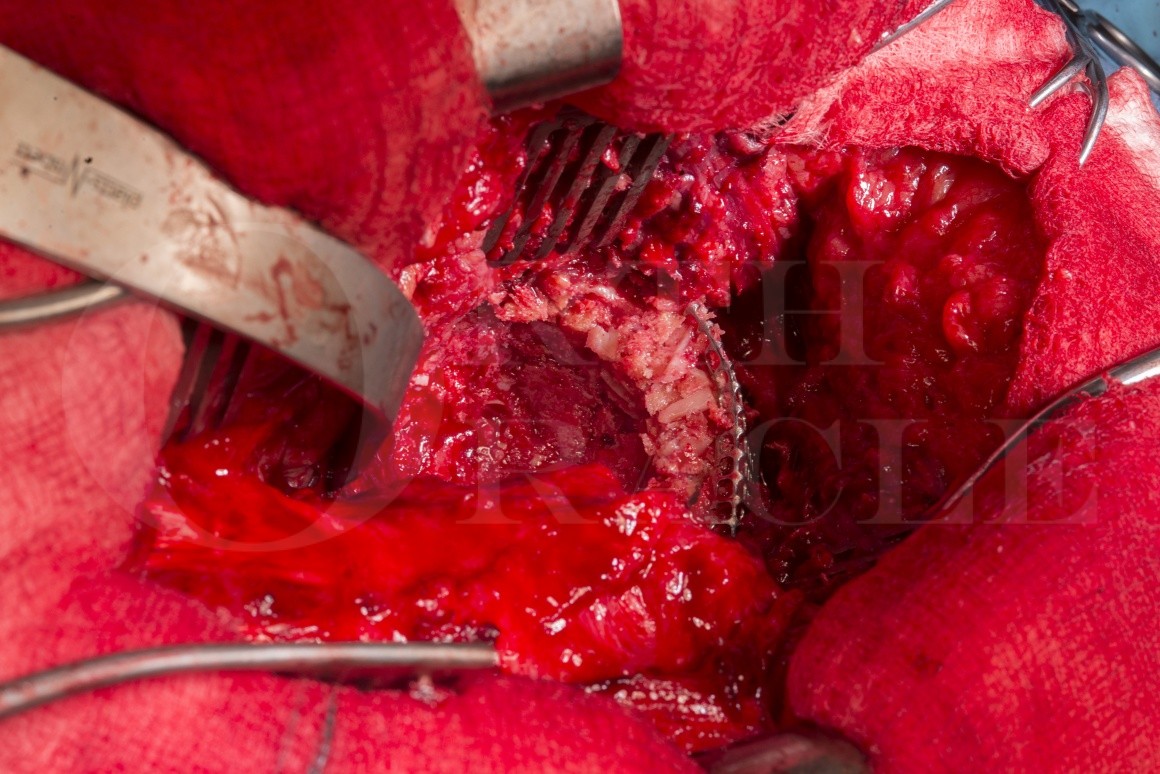
This method is used where there has been bone loss. Rim mesh is not used for constrained defects but when there has been loss of the acetabular wall (usually posterosuperior).
However rim mesh has its limits, firstly it only comes in three sizes and in my experience none is ever the right size. Also any bone loss gap that cannot be spanned by the mesh is not reconstructable using this method.
SYMPTOMS & EXAMINATION
Osteolysis may be totally asymptomatic and only picked up in routine outpatient follow-up. Some of these patients present with catastrophic failure and fracture. Rim mesh is not suitable under those circumstances.
However rather fortunately, the majority of patients will present with pain. Usually located in the groin area but pain from a loose acetabular component may be felt in the buttock region.
A standard hip examination may reveal pain in the extremes of motion, or if the cup is very loose the hip may be irritable and the patient may not allow much movement at all.
IMAGING
As ever, plain radiography is the standard. A simple AP pelvis and lateral hip usually suffice. When extensive bone loss is apparent a CT scan may be useful for assessing the amount of bone loss and confirm any suspected dissociation.
Inflammatory markers also need to be taken to rule out infection. In our unit we will use ESR, CRP and WCC.
If inflammatory markers are elevated, or even if not and the implant has loosened early (less than 10 years) we aspirate the hip for microbiology. We do not perform bone grafting in the presence of infection (although I note others have published on this).
ALTERNATIVE OPERATIVE TREATMENT
Alternatives to the rim mesh and impaction bone grafting include large uncemented acetabular components and the use of augments. An issue I have with these (as alluded to above) is that in order to get good seating of the uncemented implant it is often necessary to remove excess, normal, healthy bone. Bone preservation is paramount in the young patient in particular as they may require additional revisions in the future bone and retention of bone stock is therefore very important.
NON-OPERATIVE MANAGEMENT
If there is no risk of fracture, no significant bone loss and the patient is relatively asymptomatic there is no desperate need to revise. Lifestyle modification, acceptance of some limitations is often acceptable to the elderly frail patient versus a large potentially risky operation.
CONTRAINDICATIONS
In our unit infection would be a contraindication. Others include defects which are too large for reconstruction with rim mesh. Although I have never encountered this, patients may refuse as they do not want to receive allograft.

As is usual for a revision hip, prior planning is essential. We discuss all of our cases (even simple primaries) at a planning MDT meeting. We also have waiting list co-ordinators, and theatre team representation to discuss kit required.
The set up for this case was a ‘basic revision tray’ which contained the drills and reamers.
The Impaction bone grafting kit / X-change revision instruments are needed.
I also use the Nichol’s extractor .
Rim mesh and screws – these are made by Stryker.
I had made it clear I needed a frozen femoral head (we have an on-site bone bank which enables us to perform this operation). I accept many trusts / hospitals do not have easy access to femoral head graft and buying them in specifically for an operation can be costly.

This is the plain radiograph of a 62yr old patient. Her hip had been functioning very well for 15 years without problem until she fell of a cliff from a survivable height.
This fall had loosened a well fixed cup or had made symptomatic a loose cup. Hopefully the former. The patient had mobilised on scene, had not gone to hospital, but had ongoing groin pain. She was mobilising and remained independent. The centre of rotation of the hip was affected with medial and superior migration.
This website is not necessarily the forum to discuss the biomechanics of the hip, but it is very important, especially around exam time. There are many revision websites available etc but I would strongly recommend 2 papers that explain this field in a very straight forward and easy to understand fashion.
The radiographs show loosening in Zones I-III (DeLee and Charnley). The loosening zones, so often quoted, were published in 1976, it is a nice paper to read. A quick note when assessing ‘lines’, it is the progression of lines that is important but do not be falsely reassured if the lines appear static, also assess whether there is migration of the socket.

Anatomy & Biomechanics of the Hip. D Byrne et al. The Open Sports Medicine Journal, 2010, 4, 51-57
Basic biomechanics of the hip. D Lunn et al. Orthopaedics and Trauma, 2016, Vol.30(3), 239-246
DeLee, Charnley. Radiological demarcation of cemented sockets in total hip replacement. Clincal Orthopaedics 1976. 121:20–32

The lateral hip radiograph shows similar osteolytic lines.
As mentioned, the presence of lines is not pathognomonic of loosening, progressive lines are. Also falling off a cliff and having pain with radiolucent lines is also pretty indicative that something is wrong.

This presentation is not about the approach and dissection. We are therefore going to start with an exposed acetabular component, approached posteriorly. The femur was dislocated and the head removed.
The hip was surprisingly tight initially but sequential and progressive release of scar and capsular thinning eventually allowed a good view.

To orientate you please refer to the multiple annotations. The camera angle can make this confusing!
Ant – Anterior, Post – Posterior, Sup – Superior, Inf – Inferior, Fem – Femur, VL – vastus lateralis
The femoral head has been removed from the stem and it has been inserted into a small ‘pocket’ made anteriorly to the acetabulum. This is made with a combination of sharp and then blunt dissection.
Two important points need to be raised on this image. Firstly it is necessary to understand the position of the original acetabular rim (A). It can be seen that the implant has migrated (go back and check on the AP pelvis images). The second important point is that there is a rim defect (B). It does not look much as the cup has migrated however upon restoration of the hip centre, the true size of the defect will be made apparent.

Loose bony fragments and cement overlying the cup was removed. It was obvious that this cup was loose as firm pressure from a charnley spoon was enough to see it move. However it was still entrapped in the acetabulum with fibrous tissue and was not loose enough to simply flip out.

Enter the Nichol’s extractor. This is a great piece of kit. It consists of two parts, the poly implant extractor (shaft) (A) and the poly implant adaptor (B). The shaft has a handle and a strike plate for a hammer (C) proximally .

The distal, working end has two different diameter threads. The smaller thread screws into the polyethylene, the larger is the thread for the implant adaptor.

Prior to use, the extractor is constructed by screwing the adaptor onto the shaft. The adaptor can therefore move up and down the shaft.

A pilot hole is made in the polyethylene. It needs to be full thickness down to bone cement.

For cemented cups the hole needs to be at the apex of the acetabular component. Never make a drill hole here in uncemented cups as this usually corresponds to a hole in the shell that attaches the impaction handle. Please avoid plunging into the pelvis.

The Nicols poly extractor shaft is fully screwed into the polyethylene until it hits bone cement and the poly implant adapter is then screwed and advanced until it makes firm contact with the rim of the polyethylene cup.

The implant extractor therefore has a firm grip in the polyethylene and the adaptor component is firmly pressed against the cup rim. This forms a stable construct, movement of the handle does not result in toggling of the thread in the polyethylene.

Our extractor is made by Platts and Nisbett, a proud Sheffield company located around the corner from the hospital. I have had a brief look at other orthopaedic manufacturing companies websites whilst writing this topic and cannot find this tool anywhere. For us it is a workhorse.

Finally ensure that all soft tissue has been removed from around the acetabulum. This will be a combination of sharp dissection for overhanging soft tissue, the judicious use of osteotomes and ‘nibblers’ for overhanging cement or bone.

The extractor can then be moved up and down and side to side, breaking down the fibrous scar tissue around the implant.

This (with control) can lead to a very quick extraction.

As you can see, with a loose cup there is no further bone loss.

All of the cement is removed. In this case a quick win.

Attention is then turned to the remaining cavity. You can see the (postero)superior wall defect (A).
There are many classification systems for acetabular defects. The two most common being the AAOS classification and the Paprosky classification. The former is good at describing the anatomical defect, the latter is very good for planning surgical reconstruction. I did reference two great review articles in my earlier presentation They are relatively lengthy descriptions but in summary
AAOS Classification of Acetabular Bone Loss.
Type I – Loss of a portion of the acetabular wall or of the medial wall
Type II – Volumetric bone loss (contained) within the bony pelvis
Type III – Segmental and cavitatory bone loss
Type IV – Acetabular diastasis
Type V – Arthrodesis
Paprosky Classificaiton
Type I – Minimal bone loss, the acetabular rim is intact
Type IIa – Bone loss superiorly with limited / mild acetabular migration and an intact rim
Type IIb – There is moderate superolateral migration (<2cm)
Type IIc – Moderate medial wall lysis, mild migration but loss of the anterior column (Kohler’s line disrupted)
Type IIIa – As above but migration is greater (>2cm) and there is evidence of lysis of the posterior column (Ischium)
Type IIIb – Lysis everywhere with severe migration of the cup in an superomedial direction.
The acetabulum is full of inflammatory and scar tissue in this case.

The ‘push block’ inserts in the top of the mill and hand crank is turned.

This is removed with a combination of blunt (Charnley spoon) and sharp dissection. Always test the floor and the walls of acetablum before getting too forceful to prevent plunging into the pelvis.
The acetabulum can be divided into 4 ‘zones’ by a line drawn from the ASIS through the centre of the acetabulum. An orthagonal line is then drawn, again centred on the acetabulum. This gives a posterior-superior, an anterior-superior, an anterior-inferior and a posterior-inferior zone.
The only ‘Safe Zone’ is the posterior-superior zone. Plunging through this (with screws / drills / the charnley spoon) puts the sciatic and superior gluteal nerves at risk. The ‘Caution-Zone’ is posterior inferior. Plunging more than 20mm places the inferior gluteal nerve, the sciatic nerve, and the internal pudendal nerves and vessels at risk.
The anterior-superior ‘Danger Zone’ contains the external iliac artery and vein. The anterior-inferior zone contains the obturator nerve and vessels.

Finally, I ‘freshen up’ the bone to get active bleeding, a good bed for bone grafting. This is done with the reamer. As the defects are not usually purely hemispherical it may be that smaller reamers are needed to freshen up deeper, recessed areas. What ever size is chosen, this step should be done with caution. Palpate to confirm the thickness of the anterior and posterior walls and make a mental note from the radiographs of how thick the medial wall is. When the bone is bleeding stop. Some areas may never bleed (if really sclerotic), in these instances either the reciprocating saw or multiple small drill holes may ‘freshen’ up the bed enough, and promote bony bleeding.

You are going to have to trust me on this image. This shows the trial contemporary trial in situ. It has fallen backwards somewhat into the acetabular defect but the posterosuperior wall defect can be (partially) seen. This uncontained defect prevents reconstruction with simple cementing techniques.

This is an image of the rim-mesh that was first opened. As I stated they come in sizes of too small and too large. This falls into the former category but you can see it has a rim border (A) and then tapers out (B). This taper usually aims towards the lesser sciatic notch.

The correct size rim mesh is slid into place with a ‘tipped ball pusher’. Although not directly visible on this image the sciatic notch is supero-posteriorly. It is important that the mesh does not project into this notch as it can irritate the sciatic nerve. The nerve exits the pelvis through the greater sciatic foramen, emerging inferior to piriformis (5-10% have an early high division with the common peroneal nerve branch exiting superior to priformis). It runs on the deep surface of the superior gemellus, obturator internus, inferior gemellus and quadratus femoris, finally passing deep to the long head of biceps femoris as it enters the popleteal fossa.
If necessary, the mesh can be trimmed with sturdy scissors. Again the angles of the photograph can be misleading however the most anterior part of the mesh is just posteroinferior to the anterior inferior iliac spine.

Once in the correct position I drill pilot screw holes. I note that Stryker, in their op-tech state this is not necessary as the screws are self drilling and self tapping. However by drilling you get an accurate length measurement (instead of choosing all the screws to be 20mm) and you can easily tell if you have got an ‘air-shot’ that will give no grip. Take a look at the superior acetabular anatomy of a pelvic model. You can see that for the posterior mesh screws you need to aim the drill slightly posteriorly and inferiorly, into the posterior column and for the anterior screws the aim is inferiorly and anteriorly into the anterior column. I still get a bit twitched with these screws as there is plenty of clockwork here (see slide 20) but the columns are usually quite robust and it is usually possible (unless exceptionally scarred up) to get a finger into the sciatic notch to aid 3D spatial awareness.

The mesh is going to act in tension and therefore it only requires four or five screws to achieve stability. There will be some flexibility in the mesh at this point but impacting bone will tension it.

This image is of a freshly frozen femoral head.
Femoral heads in our institution are collected during primary hip replacement surgery. Patients are pre-screened for donation in our pre-assessment unit. There is a detailed questionnaire for patients to complete regarding risks of disease. These include patients who are at risk for blood borne viruses, ‘at risk’ lifestyles and history of cancer. In total there are 34 screening questions. Patients also have to consent to blood testing for Hepatitis B and C, HIV 1 and 2 and syphilis.
Once harvested any soft tissue is removed from the femoral head and microbiology swabs are obtained from the external cartilage. A sample of cancellous bone from the femoral neck is also sent to micro. The head is transferred to a dry sterile pot which is then frozen at -80 degrees.
On the day of surgery, before implantation but under the laminar flow, external swabs and bone samples are once again sent for microbiology.
The femoral head cannot be used directly in the bone mill as it is too large.
We do not remove any of the cartilage prior to impaction bone grafting. Retrieval studies which show that presence of cartilage cells does not effect construct stability in vivo. Outcome studies have also clinically shown no difference in outcome. Our philosophy is that removing cartilage and individually constructing large bone chips from femoral heads increases the number of femoral heads required (and cost) and increases operative time (and hence risk of infection)

The femoral head is bivalved. It needs to have defrosted sufficiently (a jug of warm Hartmans suffices). I personally hold the head in a swab but cannot recommend this to others for obvious reasons.
Slooff et al. first popularised the technique of impaction bone grafting with unwashed bone chips, although there were descriptions of using bone chips for acetabular protrusio in the mid 1970’s. There have been many variations in technique. Many have published on size and the preparation of the bone chips. Others have published on the use of a combination of allograft and synthetic bone. Ours method is but one but we have found it to be reliable and reproducible, however it does need attention to detail and practice.

Slooff T.J.et al Bone grafting in total hip replacement for acetabular protrusion. Acta Orthop Scand. 1984;55:593–596

I stop the saw before the head is completely bivalved and use an osteotome to complete the dissection. A gentle tap is usually at that is required.

As you can see in this image that the head still has a large fat / marrow content. This will be removed later. The presence of fat and marrow is thought to be important as it may affect the interlocking of bone particles and penetration of cement.
Techniques to improve the shear strength of impacted bone graft: the effect of particle size and washing of the graft. Dunlop DG, et al J Bone Joint Surg Am. 2003;85:639-46.

The structural support of the bone graft is dependent on the size of the bone chips, and the amount of impaction / ‘tightness’ of the graft. Many commercial bone mills only allow graft size of 2-5mm which in my opinion (and many others) is too small. This is why acetabular reamings should not be used. Our cutting cylinder produces 8mm chips. The bone mill is simply a glorified old fashioned mincer.

Basic science of bone impaction grafting. Schreurs BW et al Instructional Course Lectures. 2001; 50:211-20.

The femoral head fragments are inserted in the top.

This generates bone chips approximately 8mm in size.
These chips are obviously allograft and the bone is fresh frozen.
The properties of bone graft, the types of bone graft, the preparation of bone graft and healing are all very common questions in the FRCS exams.
It is important to know the properties of bone graft. Others will phrase it better but;
Osteoconductive – graft acts a framework for ingress of new bone cells
Osteoinductive – graft contains elements to induce stem cell differentiation and promote bone growth
Osteogenic – Graft contains bone cells.
Allografts, autografts, demineralised bone matrix and synthetics bone all have different properties. Graft healing also differs between cortical and cancellous graft.

Back to the case, the cutting cylinder is cleaned of residual bone.

The bone chips are washed to remove fat, blood and marrow.

They are then transferred to a swab.

I hold the neck of the swab with one hand and twist the bag of bone chips with the other, tightening the ‘sack’ squeezing out moisture.

The washed and dried bone chips.

The bone chips are transferred back to a clean receiver ready for impaction.

Bone chips are placed in the cavity and pushed to the areas of bone loss. It is important to impact the bone fully. It needs to have good interlock and be a solid construct. It should be firm to touch and not friable.

Therefore the bone chips are not added all at once, only a few at a time which are then impacted. It can be laborious and repetitive, but it is important to execute this step correctly.

We use our patented and complex osseotransferenceconveyer (formerly a desert spoon) to get the bone chips in the acetabulum.

The next two slides show the impaction bone grafting kit. It is pretty basic. The uppermost device is the sieve. This is placed against the impacted bone before using the sucker for obvious reasons.
The bottom most device is the rim impactor (of which more later).

The other pieces in the kit are increasingly sized domed impactors.

The impactors are placed against the bone chips and hit with a mallet. I personally use multiple firm taps as large force impactions risk fracture. However it is imperitive that the bone chips are firmly impacted, it does take some force. It is obvious therefore that when there is a medial defect a secured floor mesh needs inserting.
One of the most difficult areas to get good impaction is the superolateral rim. Here I ‘aim’ the impactors superiorly by dropping my hand.

Controlled impaction whilst suction is applied to the acetabulum.

As stated earlier, blood accumulating at the impaction site needs removing with the sieve. The Janker sucks through the sieve as shown.

It is important to ensure that the rim areas are firmly impacted using the rim impactor as shown. The dome impactor needs to be left in-situ when rim impacting. The graft construct should be solid and not friable, it shoud not move under pressure.
The last impactor is the same ‘size’ as the Contemporary cup. Stryker Contemporary sockets have PMMA spacers on their outer surface to aid an equal cement mantle. The acetabular cup size therefore of a Contemporary socket refers to the outer diameter of the cement spacers and therefore includes a 3 mm cement mantle.

Finally I ream the area prior to cementing with the reamer ON REVERSE. The reamer is the same size as the last impactor / proposed cup. Again pressure is maintained.

By using impaction bone grafting the posterosuperior wall defect has been reconstructed and the centre of hip rotation has been restored, improving hip biomechanics. The graft thickness should be greater than 5mm.

This is not a ‘how to do a cemented cup’ case and cemented cups are described elsewhere on the site, however…The bed of the acetabulum / graft is dried, a single mix of antibiotic loaded cement is added to the acetabulum with a cement gun (I insert cement at 1 min and 45 seconds). The cement is pressurised with a cement pressuriser (2-4mm larger than the acetabular aperture) and at 4 minutes I insert the cup. To do this is I medialise first (i.e push the cup down through the cement, with it in a ‘closed’ position) and then with pressure from the axial pusher, the cup version and inclination are dialled in. I aim for 20 degrees of anterversion and 40-45 degrees of inclination. Pressure is maintained until the cement has cured and whilst this is happening, excess cement is removed with a McDonald’s.
I would strongly recommend that readers consult the freely available ‘The Well-Cemented Total Hip Arthroplasty’ (Breucsch Malchau Eds.)
Finally I trial with femoral heads, ensuring correct tension and stability before finally impacting a new femoral head. This can be either a metal head or (occasionally) a ceramic ‘revision’ head which has a titanium alloy sleeve. A ceramic head should never be impacted on an old trunion. Increased point loading on old asperities increases the risk of ceramic fracture.

It is worth recalling the pre-op image before reviewing and contrasting the post-operative appearance. Please note that this single radiograph is slightly over-penetrated giving an appearance of femoral lysis in Gruen zone 7. This is not the case.

The postoperative radiograph reveals that the rim mesh is well held by the screws, the hip centre has been normalised, the cement has good bone penetration and the areas that were once lucent are now filled with homogeneous impacted bone.

The lateral radiograph confirms the findings of the AP as well as confirming cup anteversion.

Standard post op protocols apply.
Check bloods, thromboprophylaxis, x-ray when comfortable.
Weight bearing status depends somewhat on your confidence with the graft. Previous (historical) instructions included confined to bed for 6 weeks, followed by 6 weeks partial weight bearing and then increasing to full over 6 more weeks. This argument is that this allows greater osseointegration, however full graft healing can take up to 30 months.
I try to get all my patients full weight bearing post op. If there is to be subsidence, the final position will occur earlier. Early weight bearing has not been proven to lead to increased cup migration. The majority of patients are ‘weight bearing as tolerated.’

A general reading list for those embarking on this type of operation is as below.
Anatomy & Biomechanics of the Hip. D Byrne et al. The Open Sports Medicine Journal, 2010, 4, 51-57
Basic biomechanics of the hip. D Lunn et al. Orthopaedics and Trauma, 2016, Vol.30(3), 239-246
The Well-Cemented Total Hip Arthroplasty’ (Breucsch Malchau Eds) Springer.
DeLee, Charnley. Radiological demarcation of cemented sockets in total hip replacement. Clincal Orthopaedics 1976. 121:20–32
Slooff T.J.et al Bone grafting in total hip replacement for acetabular protrusion. Acta Orthop Scand. 1984;55:593–596
Techniques to improve the shear strength of impacted bone graft: the effect of particle size and washing of the graft. Dunlop DG, et al J Bone Joint Surg Am. 2003;85:639-46.
Basic science of bone impaction grafting. Schreurs BW et al Instructional Course Lectures. 2001; 50:211-20.

Impaction bone grafting now tends to be done for the lesser acetabular defects. TM metal, augments and custom implants have superseded the massive strut / bulk allografts that were performed more historically. This is good as outcomes from these massive procedures were poor, however these were the only tools available at the time.
However (as opposed to the TM and custom implants) there is significant long term data regarding outcomes. 15 -20 year outcomes of 92% cup survival are reported widely with survival of 79% at 20 years.
Acetabular revision with impacted morsellised cancellous bone grafting and a cemented acetabular component. Schreurs et al. 2009 JBJS


Reference

  • orthoracle.com
Dark mode powered by Night Eye