
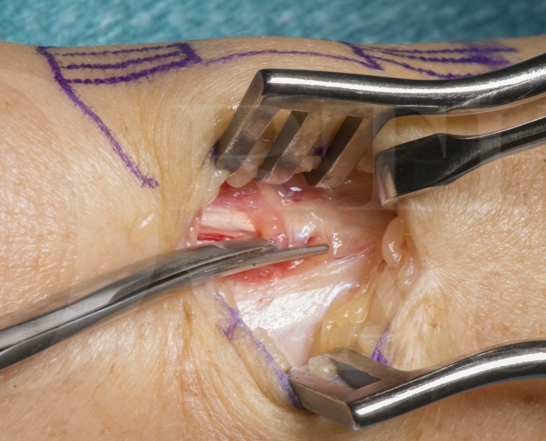
Learn the De Quervain’s release – Dorsal approach surgical technique with step by step instructions on OrthOracle. Our e-learning platform contains high resolution images and a certified CME of the De Quervain’s release – Dorsal approach surgical procedure.
This is a detailed step by step instruction through a release of the 1st Extensor compartment for a diagnosis of De Quervain’s tenosynovitis.
In 1895 Fritz de Quervain repaorted 5 cases of painful thickened first dorsal compartment of the wrist.
De Quervain’s is a non-inflammatory stenosing tenovaginitis which is often self-limiting with activity modification. The fibro-osseous tunnel or contents of the 1st extensor compartment, namely the extensor pollicis brevis (EPB) and abductor pollicis longus (APL) tendons, become thickened causing pain and possible crepitus and catching during thumb movements.
This decompression procedure is undertaken when all non-operative treatment options – activity modification, analgesia, steroid injection, splintage, physiotherapy – have been exhausted.
The surgery is very successful however it can have 2 very significant complications which create more pain and dysfunction then the original complaint and are much more difficult to resolve. These complications are volar subluxation of tendon in wrist flexion and neuropathic pain from the superficial radial nerve or lateral cutaneous nerve of the forearm branches.
Following surgery patients are put into a bulky bandage and allowed to mobilise however no heavy lifting, twisting or gripping for 6 weeks is advised.

Anatomy & Pathology
The first extensor compartment is a fibro-osseous tunnel lying on the radial styloid with a roof of the extensor retinaculum.
The compartment contains the Abductor Pollicis Longus (APL) and Extensor Pollicis Brevis (EPB) both of which can often be comprised of more than one tendon slip.
On occasion a sub (or deep) compartment against the bone can exist and must be looked for intra-operatively.
De Quervain’s syndrome is a non-inflammatory thickening of the tendons with histopathological specimens showing a thickening and myxoid degeneration, the latter being consistent with a chronic degenerative process.
Indications
Surgical decompression for De Quervains tenosynovitis is reserved for refractory cases which have persisted despite treatment with conservative measures – Activity modification, analgesia, steroid injection, hand therapy and splintage.
Surgical decompression is a last resort and although very successful in relieving symptoms the possible complications can be more painful than the initial condition, not self-limiting and more difficult to treat.
Symptoms
Patients complain of radial sided wrist pain with occasional dysthesia and pain in the distribution of the superficial radial nerve( which occurs because the nerve branches which overlie the 1st extensor compartment are irritated by the swelling). This may also effect the terminal branches of the lateral cutaneous nerve of the forearm.
Specific activities are often noted as aggravating the pain such as lifting the kettle, lifting a young child and gripping to open jars or door handles.
These activities aggravate the pain around the radial styloid due to the requirement of the EPB and APL to stabilise the thumb during pinch grip. It has also been noticed that the these muscles act as secondary stabilisers for the wrist when lifting in neutral pro-supination as with a kettle. It must be noted that an underlying diagnosis of wrist instability may be the initiating factor for De Quervain’s and thus wrist stability should be examined in these patients.
The presence of rheumatoid disease and diabetes is important to be aware due to their association with this condition.
Examination
Clinical examination starts with inspection which may be normal or may reveal swelling over the 1st extensor compartment around 1cm proximal to the tip of the radial styloid.
Evidence of previous steroid injection such a skin depigmentation or fat atrophy may also be seen over the 1st extensor compartment.
Palpation will usually elicit tenderness over the 1st extensor compartment also should also be used to exclude differential diagnoses such as tenderness over the 1st CMCJ (which may indicate arthritis).
When assessing the range of movement of the wrist particular attention should be paid to the potential existence of radial sided wrist arthritis by moving the wrist into radial deviation (not painful in De Quervains).
The range of movement of the thumb which is usually painful is flexion – taking the tip of the thumb to the 5th MCPJ – exacerbated with the addition of wrist ulnar deviation.
Special tests for De Quervains are :
Eichhoff test – Patient places thumb in palm and clenches fingers around thumb into a fist then actively ulnar deviates the wrist to elicit pain over the 1st extensor compartment.
Finklestein test – Examiner applies longitudinal traction and ulnar deviation to the thumb producing pain over the 1st extensor compartment in the direction of an ulnar deviated wrist.
WHAT test (Wrist Hyperflexion and Abduction of thumb test) – Patient hyperflexes their wrist and extends and abducts their his thumb against resistance. This test is considered to be more sensitive & specific than the Eichhoff & Finklestein tests.
Tinel’s test – can be performed by tapping over the 1st extensor compartment which can cause tingling in the distribution of the superficial radial nerve.
Other special tests that may be of use in excluding common differential diagnoses for De Quervains:
Telescoping of the wrist for 1st CMCJ arthritis, Kirk Watson for scapho-lunate instability, Kleinman shear and Reagan tests for luno-triquetral instability, Litchman midcarpal instability test.
Investigations
In the majority of cases no investigations are performed.Plain radiographs do not usually reveal any abnormality in De Quervain’s disease. However, they are useful to exclude the differential diagnoses such as 1st CMCJ or wrist arthritis or indications of wrist instability e.g. scapholunate or lunotriquetral gaps, overflexed or extended lunates.
Ultrasound may aid with diagnosis when uncertain and can be combined with steroid injection for greater accuracy of injection administration.
MRI may aid diagnosis and rule out differential diagnoses.
Non-operative treatments
For first line treatment a steroid injection is usually provided with advice on activity modification.
In addition to injection treatment review with a hand therapist can be provided for stretching and strengthening exercises and a supportive splint for certain activities which are unavoidable can be provided.
Simple analgesia such as paracetamol and non-steroidal anti-inflammatory medications can help with symptoms.
There are no alternative operative treatments .

The surgery can be performed under local, regional or general anaesthetic.
The patient is supine with their arm on an arm board and upper arm tourniquet inflated to 250mmHg.
An assistant or lead hand is useful to support the hand during the surgery.
Otherwise no specialist equipment is required.


The surgery is performed as a day case procedure and patients, who are in a bulky bandage are allowed to mobilise however no heavy lifting, twisting or gripping is permtted for 6 weeks.
Patients are review at 1 week to change the dressing and assess the wound and if stiff or apprehensive at this point can be seen by the hand therapy team for advice.
At 6 weeks patients are expected to return to most activities and work.

Complications:
Superficial infection <1%
Nerve injury/painful scar 4% – often caused over-zealous retracting no the nerve branches seen within the wound. Nerve injury may also be cause by transection creating a neuroma or the scar tethering the nerve branches causing a tender scar or traction nerve pains.
Persistent symptoms – suggests incomplete release or incorrect diagnosis.
Tendon dislocation – rare. Volar subluxation of tendons in wrist flexion caused by over excision of sheath, release of volar rather than dorsal attachment of compartment or excessive early loaded wrist flexion.
Chronic regional pain syndrome – rare.
Papers of interest:
Hand therapy versus corticosteroid injections in the treatment of de Quervain’s disease: A systematic review and meta-analysis. Cavaleri R, Schabrun SM, Te M, Chipchase LSJ Hand Ther. 2016 Jan-Mar;29(1):3-11.
A systematic review to assess hand therapy versus injection for the treatment of De Quervain’s. This study revealed that both corticosteroid injections and hand therapy improved pain and function from baseline. No significant difference was seen in successful outcomes between groups however there was a significant increase in success when a combine approach of orthosis and corticosteroid were compared with the treatments alone.
Patient satisfaction and outcomes of surgery for de Quervain’s tenosynovitis. Ta KT1, Eidelman D, Thomson JG. J Hand Surg Am. 1999 Sep;24(5):1071-7.
A retrospective study of 43 patients with mean 3 year follow up post decompression. 5% recurrence, 2% superficial radial nerve injury, 2% with severe scar pain. 91% complete resolution with no complications.
Long term results of surgical release of de Quervain’s stenosing tenosynovitis. Scheller A1, Schuh R, Hönle W, Schuh A. Int Orthop. 2009 Oct;33(5):1301-3.
Papers of interest:
Hand therapy versus corticosteroid injections in the treatment of de Quervain’s disease: A systematic review and meta-analysis. Cavaleri R, Schabrun SM, Te M, Chipchase LSJ Hand Ther. 2016 Jan-Mar;29(1):3-11.
A systematic review to assess hand therapy versus injection for the treatment of De Quervain’s. This study revealed that both corticosteroid injections and hand therapy improved pain and function from baseline. No significant difference was seen in successful outcomes between groups however there was a significant increase in success when a combine approach of orthosis and corticosteroid were compared with the treatments alone.
A systematic review to assess hand therapy versus injection for the treatment of De Quervain’s. This study revealed that both corticosteroid injections and hand therapy improved pain and function from baseline. No significant difference was seen in successful outcomes between groups however there was a significant increase in success when a combine approach of orthosis and corticosteroid were compared with the treatments alone.
Case series of 94 patients following surgical decompression under local anaesthetic with longitudinal incision and partial resection (<3mm) of sheath. 80 female 14 male. Mean age was late forties. Outcomes included 1% superficial wound infection, 1% delayed wound healing, 4% transient SRN lesions. 94% successful release without complications.


