
Learn the Digital nerve neurolysis plus Vivosorb barrier wrap (Polyganics) and Z-plasties to scar contracture left little finger surgical technique with step by step instructions on OrthOracle. Our e-learning platform contains high resolution images and a certified CME of the Digital nerve neurolysis plus Vivosorb barrier wrap (Polyganics) and Z-plasties to scar contracture left little finger surgical procedure.
Complications of digital nerve repair include neuroma-in-continuity formation and tether in scar. A neuroma is clinically defined by a non-progressive Tinel’s sign at the site of injury and repair with no functional sensory recovery distal to the repair. Nerve tether is typified by a painful scar and exacerbation of symptoms of pain and paraesthesiae on passive stretch.
When there is no distal sensory function consideration should be given to revision surgery for excision of neuroma and graft reconstruction of the resultant gap. In such cases the use of a processed nerve allograft is superior to autograft when the patient has significant neuropathic pain and sensitisation. If there is some distal protective sensation and the Tinel’s sign is moderate t the site of repair, the surgeon should consider a neurolysis at the site of repair and the option of wrapping the repair site with a barrier to further scar formation.
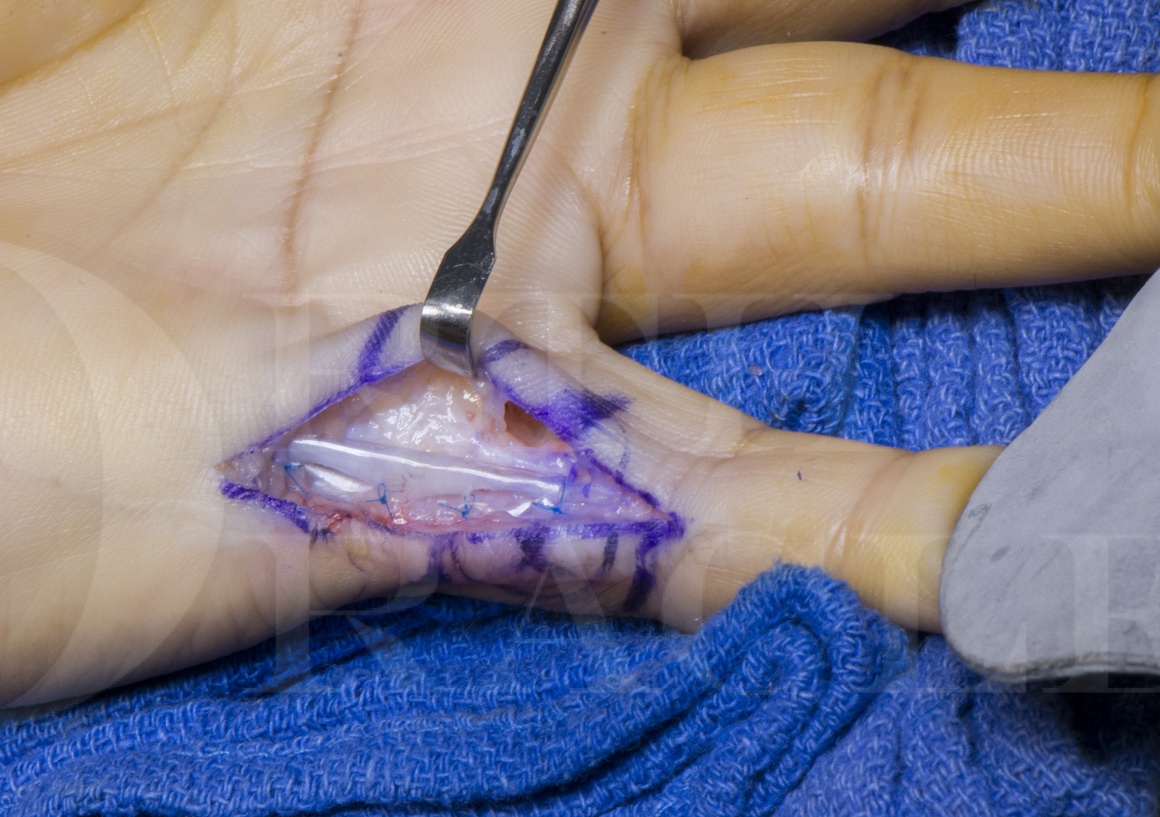
The Vivosorb is a polycaprolactone bioresorbable membrane that can be wrapped around the nerve following neurolysis and protects the nerve from scar formation in the weeks following revision surgery.

INDICATIONS
The indications for neurolysis include a tethered nerve in scar following repair with passive extension pain, with some sensory recovery in the distal territory supplied by that digital nerve.
SYMPTOMS & EXAMINATION
A tight or tethered scar crossing flexor creases following primary surgery and repair of a digital nerve can be released and lengthened with multiple Z-plasties. There is a Tinel’s sign (pain and dysaesthesiae in the territory of the nerve elicited by gently tapping over the repair site). Distal sensory recovery should be sufficient for at least diminished protective sensation (Semmes-Weinstein monofilament pressure thresholds of 2g). Less than this may suggest that excision of the neuroma and reconstruction of the resultant gap may be required. Stretching of the scar produces pain (neurostenalgia) due to nerve tether at the repair site.
IMAGING
Ultrasound may demonstrate neuroma at the repair site but is not essential because the diagnosis may be made with a thorough systematic clinical examination.
ALTERNATIVE OPERATIVE TREATMENT
The area can be explored and if a large neuroma in continuity is identified without good distal sensory recovery then the neuroma can be excised and the resultant gap reconstructed with reversed autologous sensory nerve graft or with a processed nerve allograft. I prefer the use of an allograft in such situations because it avoids the risk of creating a sensitised donor nerve site in an already sensitised individual with neuropathic pain. If the scar is extensive and not amenable to release and Z-plasties, resurfacing of the area may be indicated with a pedicled or free flap.
NON-OPERATIVE MANAGEMENT
Before contemplating surgery the patient should have a prolonged period of rehabilitation with a hand therapist with at least 3 months of therapy targeted at scar maturation, mobilisation and desensitisation. Silicone gel massage and night extension thermoplastic splinting may encourage scar remodelling. Sensory re-education should be aimed at improving the quality of the distal sensation following repair (tactile gnosis).
CONTRAINDICATIONS
Contra-indications to surgery include an unstable scar, and infection. A history of contamination and infection at the original surgery is a relative contra-indication to using a synthetic barrier wrap around the nerve at the time fo revision surgery.

The surgery is performed under regional anaesthesia with prophylactic antibiotics in case of nerve barrier application and an upper arm tourniquet is used to achieve a bloodless field. A lead hand supports the digit during dissection. Surgical rubber loops are used to identify the nerves and to provide gentle traction during neurolysis. Microsurgical instruments are recommended to complete the neurolysis and to position and suture the Vivosorb barrier wrap.


The limb is elevated to reduce post-operative swelling, reduce pain and improved wound healing.
The dressing is reduced at 1one week post-operatively and the wound inspected and redressed with a light dressing.
A volar hand-based thermoplastic splint is made and moulded to the hand and ulnar 2 digits to maintain extension posture of the finger and prevent scar contracture.
The hand is mobilised at this stage but the splint is applied intermittently during the day for rest and full-time at night for 6 weeks.
The hand is left free from splintage during the day at 2 weeks when the sutures are removed.
At this stage range of motion exercises are commenced with hand therapy.
Scar management strategies including moisturising, massage and desensitisation are commenced from three weeks post-operatively.

The Vivosorb is a useful adjunct for wrapping scarred nerves after neurolysis, particularly where there is scar involving the epineurium. It is made of a PLC bioresorbable polymer and as such is more acceptable to some patients than alternatives including the AxoGuard nerve protector which is made from a loosely layered porcine collagen extra-cellular matrix.
The Vivosorb has little published on its performance in vivo, however the material has been studied widely as a conduit (Neurolac™) in bridging nerve defects.
Publications:
Chiriac S, Facca S, Diaconu M, Gouzou S, Liverneaux P. Experience of using the bioresorbable copolyester poly(DL-lactide-ε-caprolactone) nerve conduit guide Neurolac™ for nerve repair in peripheral nerve defects: report on a series of 28 lesions.J Hand Surg Europ. 2012 May;37(4):342-9. doi: 10.1177/1753193411422685. Epub 2011 Oct 10.
This paper comments on the advantages of the Neurolac including transparency, bioresorption and semi-permeability as a conduit in nerve repair.
Reference
- orthoracle.com


