
Learn the Digital nerve repair: Conduit-assisted repair with Neurolac (Polyganics) surgical technique with step by step instructions on OrthOracle. Our e-learning platform contains high resolution images and a certified CME of the Digital nerve repair: Conduit-assisted repair with Neurolac (Polyganics) surgical procedure.
Digital nerve repairs are common operations in hand trauma. It is important to suspect digital nerve injury in a hand laceration through a sensory assessment. Basic microsurgical skills are required for performing a digital nerve repair. These operations can be prepared using a loupe magnification or under microscope.
Digital nerve injuries can occur at various levels. A practical way to describe them is to use the zones of the flexor tendons injury such as zone 1 to 3. Alternatively the surface landmarks such as PIP joint level or DIP joint levels can be used. Digital nerve repairs are technically challenging beyond their trifurcation at the DIP joint level and most people do not attempt repair beyond this level.
Certain injuries such as circular saw injuries can result in loss of a segment of the digital nerve. In these cases the options are either not to repair the nerve and bury the stump proximally in the muscles in the palm, use an autologous nerve graft(either posterior interosseous nerve or medial cutaneous nerve of the forearm), allograft or a nerve conduit. A nerve conduit as the one used in this case can bridge gaps up to 20mm.
Digital nerve injuries can occur along with tendon and digital vessel injuries. Assessment of circulation through capillary refill time(normally less than 2sec), temperature and pulp turgor can detect vascular injury requiring repair. Tendon injuries are assessed by testing the FDS and FDP tendons separately. FDPs can be tested by asking the patient to flex the DIP joint whereas FDS tendons are tested by immobilising the DIP joints of the uninjured joints in extension and asking the patient to flex the finger. If the patient has a vascular injury needing repair, the surgery is performed urgently and the procedure is called ‘Revascularisation of the finger’. At least one digital artery is repaired and where there is a segmental loss, a vein graft is used to reconstruct the artery. Flexor tendons are repaired as per the preference of the surgeon and the current trend is towards a four-strand core suture and epitendinous sutures.
When a patient has a combined flexor tendon and digital nerve injury, flexor tendon repair is done first followed by digital nerve repair.
The repair performed here is done using a synthetic neurotube as a protective layer over the repair. This however is a newer advance in nerve repairs with the aim of reducing the risk of a neuroma and a sensitive scar. Neurotubes such as these are either made of collagen or synthetic polymers. The one used here is made of Polycaprolactone(Neurolac, Polyganics Ltd). These tubes can also be used as conduits for bridging nerve gaps up to 20mm.

INDICATIONS: The indication for repairing a digital nerve is most commonly a traumatic laceration. Sharp injuries such as knife and glass are the common ones. Rarely a digital nerve requires repair following resection of a tumour involving the nerve but this often requires a nerve graft.
SYMPTOMS & ASSESSMENT: Patients seldom volunteer numbness as a symptom following a digital nerve injury. It is therefore important that the sensations are formally assessed while examining a hand injury. Crude sensations are often preserved even when the nerve is completely transected due to the deeper proprioceptive receptors from the bones and joints. Two point discrimination and Tens test(asking the patient to give a numerical score to sensation, given that the normal contra-lateral digit is 10) are useful test. As a bare minimum one should compare the sensations with an uninjured digit.
INVESTIGATION: Investigations are not required to diagnose a digital nerve injury. X-Rays are indicated if an underlying fracture or foreign body is suspected.
OPERATIVE ALTERNATIVES: Digital nerve repairs are usually performed without the additional protection afforded by entubulation. There are a number of commercially available devices for nerve entubulation made from bioresorbable polymers or from collagen. An alternative biological device is an autologous vein with can be positioned over the nerve co-aptation site. Conduits are designed to bridge small gaps without the need for nerve grafting. The addition of a conduit to a microsurgical nerve repair may confer advantages in terms of reducing axon escape at the repair site, reducing extrinsic scar compression or tether at the repair site and additional support of the repair site may allow the repair to be completed with less handling and suture distortion of the nerve ends resulting in less intrinsic scar formation.
NON-OPERATIVE ALTERNATIVES: There are no alternatives to treatment apart from repairing the nerve. However a non-critical digital nerve can be treated without repair when there is a segmental loss and a nerve graft is required. In these cases, there is a risk of an neuroma formation which may be symptomatic.
CONTRAINDICATIONS: When there is a heavily contaminated wound, digital nerve repair is deferred till the wound is debrided and any risk of infection reduced.

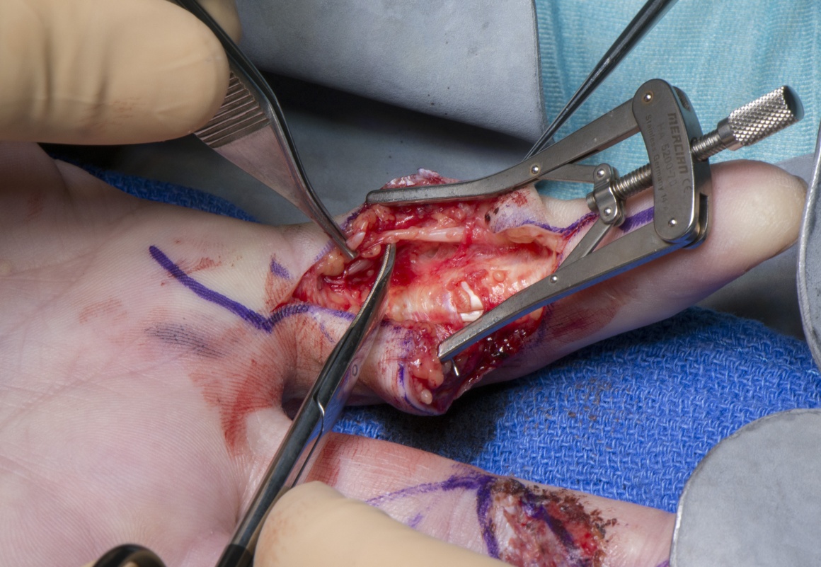
Surgery can be performed under a General or Regional anaesthesia. In this case, it is being performed under a Brachial block. The arm is cleaned and prepped. A single dose of intraveous antibiotic is administered. An upper arm tourniquet is inflated to a pressure of 250mm Hg. The hand is positioned in a Lead hand with towels underneath as shown. As can be seen in the photograph, there are two irregular lacerations over the volar aspect of the index and middle fingers. They were sustained through circular saw injuries. The edges of the wound are therefore ragged.
In the case shown here there was an injury to the ulnar side digital nerve of the index finger.


Patients are advised to keep the hand elevated. They are discharged home the same day with oral analgesics.
The first dressing change is usually done between 5-7 days. Patients are advised to start mobilisation from the first week. Splints are not necessary for digital nerve repairs done without tension.
Sutures are removed between 10-14 days.
Hand therapy exercised are continued and the patients reviewed every six weeks monitoring nerve recovery. An advancing Tinel’s sign is a sign of nerve recovery.
The time taken for recovery depends on the distance of the injury from the tip of the finger. For example an injury 10cm proximal to the finger tip may take upto 3 months for recovery.
The results of digital nerve repair are variable depending on the type of injury(clean cut injuries recover better), age of the patient(young patients recover better), location of injury(distal injuries recover faster) and the quality of repair(repairs done using microscope have a better prognosis).
Digital nerve repairs can be complicted by neuroma formation and neuropathic pain in the post-operative period. If these do not settle down, revision surgery through neuroma resection of sheathing the nerve with a biological or synthetic membrane may be beneficial.

Slutsky DJ. The management of digital nerve injuries. J Hand Surg Am. 2014 Jun;39(6):1208-15.
2. Bertleff MJ, Meek MF, Nicolai JP.
A prospective clinical evaluation of biodegradable neurolac nerve guides for sensory nerve repair in the hand. J Hand Surg Am. 2005 May;30(3):513-8.
Reference
- orthoracle.com


