/

Technique: EIP to EPL Tendon Transfer

Learn the EIP to EPL Tendon Transfer surgical technique with step by step instructions on OrthOracle. Our e-learning platform contains high resolution images and a certified CME of the EIP to EPL Tendon Transfer surgical procedure.
The extensor pollicis longus (EPL) tendon extends the thumb interphalangeal joint and also contributes to metacarpophalangeal joint extension. It serves an important role in hand opening and fine control of thumb position.
Although an acute injury resulting in division of the tendon will merit direct repair, many other causes of EPL dysfunction may be treated by tendon transfer. The commonest indication for EIP to EPL transfer is following attrition rupture of the EPL in the 3rd extensor compartment of the wrist associated with a distal radius fracture.
The commonest and certainly the simplest donor tendon to transfer to EPL is the extensor indicis proprius tendon in such cases.


INDICATIONS:
The patient in this case had a spontaneous rupture of the EPL tendon following a distal radius fracture that was treated non-operatively in a plaster cast. This is a common mechanism of injury and is thought to result from attrition rupture of the tendon secondary to extravasation of blood and possibly fracture debris into a tight third extensor compartment. A watershed zone within the intrinsic blood supply to the EPL tendon is also thought to be a contributing factor.
Other causes of a closed rupture may include inflammatory arthropathy such as rheumatoid arthritis or other forms of synovitis or tenosynovitis. Such ruptures have also been attributed to steroid therapy, excessive abnormal wrist motion, or a bony spur at the distal radius or scaphoid.
An open laceration of EPL may also require EIP to EPL tendon transfer if for example, presentation is delayed and primary repair is not possible or in cases where primary repair has failed.
It is important to note that a lack of EPL function as part of a non-recovering radial nerve palsy will usually also result in EDC and EIP dysfunction. Various alternative surgical options are described elsewhere for radial nerve palsy.
SYMPTOMS & ASSESSMENT:
The patient will usually present with weakness in thumb MCP extension and a lack of IPJ extension.
This may be accompanied by acute pain, particularly at the time of rupture and is typically felt over Listers tubercle. Pain is often not a predominant feature in spontaneous rupture at the time of presentation.
The clinical examination must include a test to establish the presence of EDC and EIP to the index finger
INVESTIGATION:
In cases where the diagnosis is in doubt, imaging using ultrasound may be useful. This can also help establish discontinuity in EPL as well as presence of the donor tendon. It may also demonstrate synovitis or tenosynovitis.
X-ray can be used to demonstrate the presence of a bony spur or other degenerative change.
OPERATIVE ALTERNATIVES:
As mentioned above, direct repair or interposition graft may be suitable options in select cases. Equally, some patients may be better served by an arthrodesis.
NON-OPERATIVE ALTERNATIVES
In patients that are not suitable for any form of surgery, functional outcome is likely to be poor. In very low demand patients one may consider non-operative management. Often an occupational therapist may be able to assist with a custom made splint.
CONTRAINDICATIONS:
Caution is advised operating on those patients with poor skin quality or poor vascularity. Poor pre-existing thumb function, for example secondary to advanced arthritic disease of the IPJ may mean that a tendon transfer does not have the desired effect. It is, as ever important to assess each patient as an individual and discuss the risks and benefits of surgery with him/her.

The patient is appropriately anaesthetised using either general anaesthesia or a regional anaesthetic block technique.
The patient is positioned supine with the limb positioned on an arm table. An upper arm tourniquet is used and inflated prior to commencing the procedure.
Three incisions are marked out. A 1cm transverse incision over the index MCPJ, a 1-2cm transverse incision overlying the EIP just distal to the extensor retinaculum, and finally a 2-3 cm longitudinal incision over the EPL tendon at the thumb metacarpal.
Tendon instruments and bipolar diathermy are required.

With the patient anaesthetised one may now freely perform a tenodesis test to confirm discontinuity of the EPL tendon.
With the wrist flexed, the thumb IPJ would ordinarily undergo passive extension due to the effect of an intact EPL. Here we demonstrate a lack of normal tenodesis at the thumb.
One can also see the index finger demonstrating a normal tenodesis effect.

The hand is placed upon a rolled up towel with thumb supported and the wrist slightly extended at approximately 30 degrees.

Once the incisions have been carefully marked out and the tourniquet inflated, the first incision is made over the thumb MCPJ.
Skin and fascia are divided sharply. One may occasionally see a branch of the superficial radial nerve here and therefore it is important to look for any nerve branches and protect them.
The two extensor tendons to the index finger will be seen at this level. It is important to remember that the more ulnar sided tendon is the EIP.

This ulnar sided EIP tendon may then be dissected free from surrounding subcutaneous tissues at this level and placed into a tendon hook.

Gentle traction of EIP will demonstrate its anatomical position just distal to the retinaculum and it is here that the second incision must be centred.

The EIP tendon is identified and freed up from surrounding structures.
Gentle traction on this tendon through both the proximal and distal incisions confirms that the correct tendon has been identified prior to division. One should also visualise the index EDC tendon and ensure this is intact before dividing the EIP.

A pair of Carroll tendon forceps are now passed from the EPL side and the EIP tendon is grasped and pulled through the tunnel and delivered to the thumb wound.

A blade is now used to divide the EIP transversely at the distal incision. Care is taken to protect the index EDC tendon.

The tendon may now be pulled out of the proximal incision.
This should not require any excessive tension to be exerted. If the tendon does not emerge easily, it may be require further freeing from surrounding subcutaneous tissues in between the two incisons. Excessive pulling risks damaging the tendon or even avulsing its muscle belly.

The EIP tendon should be wrapped in damp gauze to prevent dessication.
A third, longitudinal incision is now made over the thumb metacarpal.

Care is taken to protect any superficial radial nerve branches that may be seen here.
The dissection is continued down to the EPL tendon which often lacks the tension seen in normal tendon. It may appear bunched up here.
The proximal end is not visualised. It is often retracted proximally into the forearm and does not require exposure.

Once the EPL is identified, one must now develop the tunnel through which the EIP will be passed.
This is in the subcutaneous plane and is made by blunt dissection between the two incisions. It is important to centre the tunnel over the site of eventual tendon coaptation to ensure a straight line of pull. The tunnel should be deep to cutaneous nerve branches to prevent damage during tunnel creation or irritation by the tendon following the reconstruction.


It is important to ensure there are no adhesions or soft tissue interposition in the tunnel that may prevent free gliding of the EIP donor tendon.

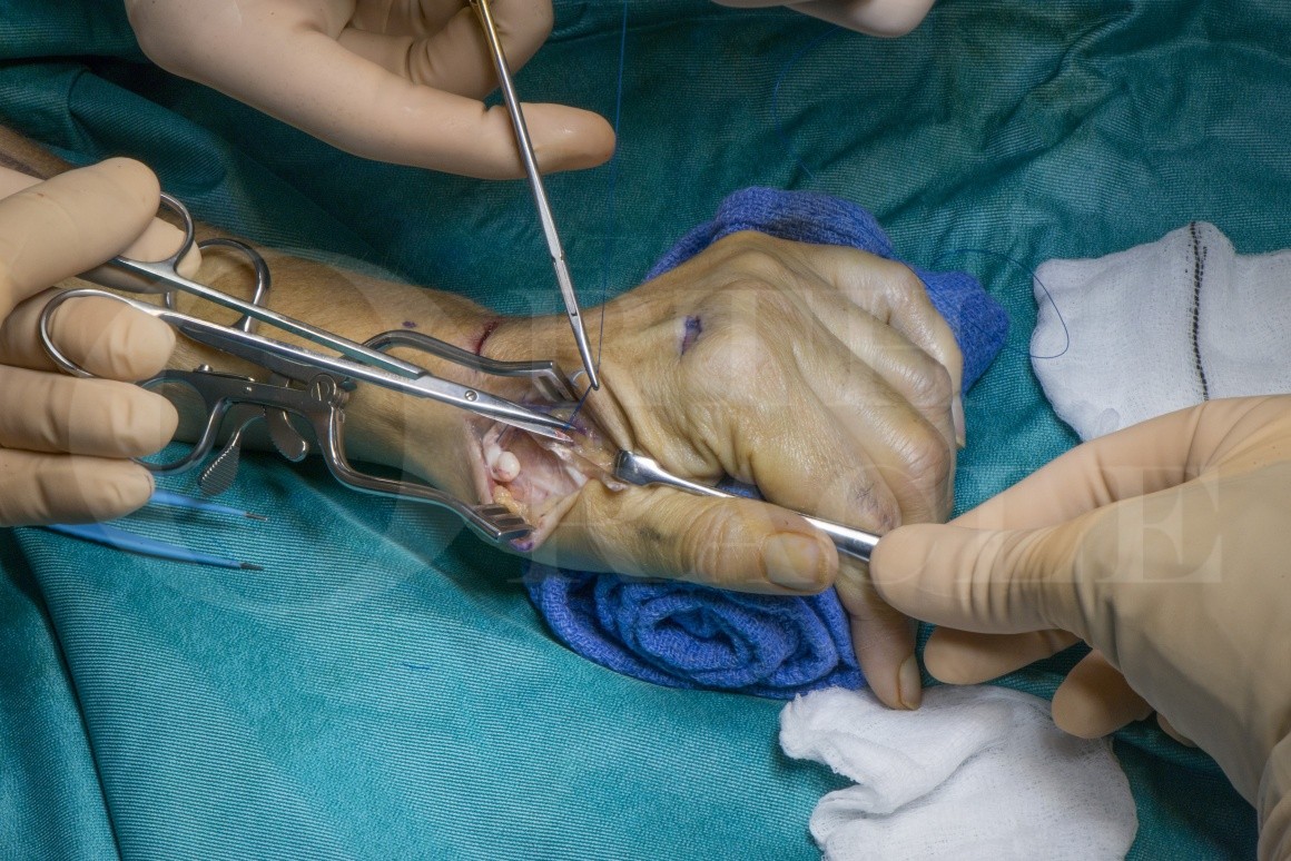
A tendon weaver is now used to penetrate the EPL tendon safely, in a radial to ulnar direction, at the ideal point for EIP insertion. This point must be carefully determined by placing the two tendons together with the wrist and thumb in the desired position.
The wrist is positioned in 30 degrees of extension, with the thumb MCPJ in neutral. The IPJ and trapezio-metacarpal joint should be fully extended.
The EIP tendon is grasped and pulled through.

With this initial tendon pass, the tension can be set prior to inserting the first holding suture.


The wrist is maintained in an position of 30 degrees of extension, with the thumb MCPJ in neutral. The IPJ and trapezio-metacarpal joint should be fully extended.
Tensioning is a matter of judgement but slight over-tension as described here is recommended as some tension will often be lost during the rehabilitation phase.

The rolled towel and an assistant are required to hold the wrist and thumb in position whilst the first suture is passed.
The weaves are sutures using 3/0 non-absorbable suture. A monofilament such as Prolene is used here.

A Pulvertaft weave technique may be used but in some patients this results in a bulky tendon transfer.
An alternative is to use a single weave followed by a side to side apposition using multiple cross-stitch sutures. This has been demonstrated to have superior strength and better gliding characteristics.

With the transfer secure, one may now cautiously flex and extend the wrist a few degrees to demonstrate adequate gliding of the tendon transfer.
As the wrist is extended beyond 30 degrees, the thumb IPJ begins to flex into neutral demonstrating that tension should be adequate to allow flexion and extension of the thumb.

After careful haemostasis, and irrigation of all wounds, the wounds are closed.
If there are any concerns about problematic bleeding it is worth releasing the tourniquet at this stage and performing a careful haemostasis.
It is important to maintain thumb and wrist position as described above during these steps in order to protect the tendon transfer until the plaster is applied.

A few interrupted deep sutures with a 3/0 vicryl may be required to bring the edges together.
A 3/0 monocryl subcuticular running suture is used for skin closure.
The wounds are supported with steristrips.

Finally, dressings are applied. Adhesive dressings are covered with a roll of wool. The thumb and wrist are supported in a volar plaster slab with the wrist in 30 degrees of extension and the CMCJ and IPJ fully extended. The MCPJ should remain in neutral.

The patients wounds are reviewed at 1 week and a splint can be fashioned at this stage to hold the thumb in extension and abduction with the wrist extended at 30 degrees.
At 4 weeks the patient should commence gentle thumb mobilisation exercises with intermittent splintage, and at 6 weeks the splint may be discarded.
A good hand therapist is essential to guide and tweak this rehabilitation process as appropriate for individual patients.

Excellent results have been widely reported. In 1997 De Smet followed up 13 patients and found good or excellent results in 12. There was often an extension lag at the thumb IPJ and MCPJ. Dexterity and grip force were excellent. Extension force at the index finger was reduced to 65% of the contralateral side.
Acta Orthop Belg. 1997 Sep;63(3):178-81.
The procedure is more reliable at restoring thumb extension than tendon grafting, direct repair or other types of tendon transfer.
Hand Clin. 1995 Aug;11(3):411-22
Biomechanical studies show that the moment arm and mechanical properties of the EIP transfer via a subcutaneous tunnel closely resemble that of the EPL.
J Hand Surg Am. 2003 Jul;28(4):661-8
Jan Friden and colleagues have used a cadaveric model to demonstrate stronger and stiffer tendon coaptation using a single tendon pass with a side to side suture technique, when compared to the traditional Pulvertaft weave. This may allow earlier loading with fewer adhesions.
J Hand Surg Am. 2010 Apr; 35(4): 540–545.


Reference

  • orthoracle.com
Dark mode powered by Night Eye