

Learn the Total wrist fusion for Volkmann’s ischaemic contracture (Using Synthes fusion plate) surgical technique with step by step instructions on OrthOracle. Our e-learning platform contains high resolution images and a certified CME of the Total wrist fusion for Volkmann’s ischaemic contracture (Using Synthes fusion plate) surgical procedure.
Volkmann’s ischemic contracture is a complication of compartment syndrome of the forearm. Untreated compartment syndrome results in degeneration of the forearm muscles and nerves resulting in contractures.
It is classified by Holden into Type I and II. In Type the pathology is proximal to the forearm such as a vascular occlusion or injury.
Type I can be mild, moderate or severe.
In the mild type, there is involvement of the deep muscle compartment mainly FDPs to middle and ring fingers. There may be partial involvement of FPL and pronator teres muscles and median nerve.
The moderate type is called the classic type. In this there is involvement of the FDPs, FPL and pronator teres. Median nerve is involved and there may be partial involvement of the ulnar nerve. The hand is in an ‘intrinsic minus’ posture.
In the severe type all flexors and pronators are involved with partial or total involvement of the extensors and instrinsics. Median and ulnar nerves are involved. The hand is in an intrinsic minus posture. Median and ulnar nerves are involved. Soft tissue coverage may be tight.
In Type II the muscle injury is limited to the site of direct trauma. The extent of muscle injury is usually limited to the forearm compartment. There may be damage to the nerves and loss of soft tissue. The extent of the finger deformity is related to the degree of muscle injury.
Corrective surgery for Volkmann’s ischaemic contracture is dependent on the type of contracture and degree of involvement of various muscle compartments. The case discussed here is a Holden Type I severe type from a vascular occlusion in the upper arm. He presented with an acute compartment syndrome which was released but unfortunately it was delayed and there was already muscle necrosis.
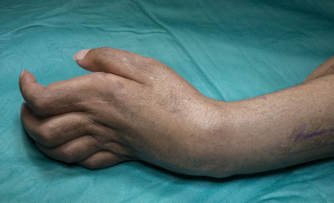
He presented with a fixed flexion deformity of the wrist, hypertension at MCP joints of the fingers and flexion at interphalangeal joints. An MCP joint release using dorsal capsulotomy was done initially and a flexor muscle slide was attempted later. Unfortunately both these operations were unsuccessful.
This operation is aimed at correcting the wrist deformity through a total wrist fusion and a later tendon transfer to move the ECRL to FDPs of the fingers. He may also need a fusion of the thumb CMC joint in an anatomical position and a transfer of EIP to FPL.
The steps of a total wrist fusion is explained through this operation.
When performing a total wrist fusion, the radio-carpal joint, capito-lunate joint and third CMC joint are fused. In this case the joints between capitate and hamate and between triquetrum and hamate are also included in the fusion to increase the strength. The distal radio-ulnar joint is left intact which usefully preserves pronation & supination.

INDICATIONS:
Wrist fusion is an operation performed for several indications including bony and soft tissue problems.
It is commonly performed for advanced arthritis involving the radiocarpal and mid-carpal joints. Alternatively for isolated radiocarpal joint arthritis, a radio-scapholunate fusion can be performed and likewise for arthritis between the carpal bones, limited wrist fusions can be performed.
It is also performed as a salvage operation after a failed limited fusion such as a four-corner fusion or failed wrist arthroplasty.
It can be an option for patients with brachial plexus or high peripheral nerve injury where there are no tendons working to move the wrist or to use the wrist tendons for providing finger movements after fusing the wrist.
The indication to fuse the wrist in a Volkmann’s ischaemic contracture is when there are no suitable soft tissue operations to improve the wrist position.
SYMPTOMS & EXAMINATION:
Patients for wrist fusion are usually those with pain and limited movements from arthritis.
In this case, the patient has a fixed flexion deformity of the wrist, hyperextension of the MCP joints and flexion deformity of the IP joints. He has imparied sensations of the median and ulnar nerves. His problems are related to soft tissues and the bony anatomy is normal.
IMAGING :
While assessing for wrist fusion in case of bony problems imaging is indicated pre-operatively. In most cases plain X-Rays in AP and Lateral views are sufficient. However if the joint surfaces cannot be assessed accurately in less severe arthritis, CT scan is useful. MRI scans are usually not needed.
X-Rays were performed in this case and were normal.
ALTERNATIVE OPERATIVE TREATMENT:
There are several techniques for wrist fusion and some techniques are preferred depending on the indications and surgeon’s preference. The common methods are using plate and screws, Steinmann pin inserted through the head of the third metacarpal head or cross K-wires. The latter two are preferred in Rheumatoid arthritis patients where the bone stock is poor. In this case we opted to use a plate and screw fixation as he is a young fit individual with good bone stock and we can position the wrist in slight extension which will help to relax the extensor tendons of his fingers thereby improving the finger flexion.
In general the alternatives to a total wrist fusion, where it is performed for arthritis are wrist replacement or denervation procedures. Replacement of the wrist joint is being increasingly performed. Wrist denervation involves dividing the anterior and posterior interosseous nerves as well as any other smaller nerve branches supplying the joint.
The alternative procedure in this case would be a proximal row carpectomy but with the attenuation of the wrist extensor tendons this would provide a less reliable functional outcome.
NON-OPERATIVE MANAGEMENT: For patients with arthritis, there is an option of conservative treatment using splints, analgesics or steroid injections. This patient has significant functional impairment due to the poor position of his wrist and therefore conservative options do not exist.
CONTRAINDICATIONS: Active infection in the skin or soft tissues is an absolute contra-indication for wrist fusion. Relative contra-indications include poor bone stock and a non-compliant patient.

The surgery is being performed under a brachial block with an upper arm tourniquet. A single dose of 1gm Flucloxacillin has been administered intravenously. The hand is positioned on the hand table and prepped and draped. The typical deformities of Volkmann’s ischemic contracture can be seen with the wrist flexion and clawing of fingers.


Patients is kept overnight for hand elevation and analgesia.
After discharge, patient is reviewed in the dressing clinic in one week.
Dressing are removed and a simple Mepore dressing applied.
A below elbow POP is applied upto the MCP joints. Hand therapy is started to mobilise the Thumb and fingers actively and passively. This is kept for another 5 weeks and a check X-Ray is performed. If it is satisfactory POP is discontinued and hand therapy continued.
Patients are advised that they can start light activities after coming out of the POP in six weeks. If there is satisfactory bony union on X-Rays, they can start strengthening exercises as six weeks and by three months can do moderate physical activity. By six months, the fusion is usually strong enough for all physical activities.
After a total wrist fusion patients can perform most day today activities including driving. Patients learn to adapt for loss of wrist movements for most activities. However sports activities such as racquet sports and swimming are difficulty due to limitation of wrist movements.

1.Houshian S, Schrøder HA. Wrist arthrodesis with the AO titanium wrist fusion plate: a consecutive series of 42 cases. J Hand Surg Br. 2001 Aug;26(4):355-9.
The authors have looked at 42 cases of total wrist fusions. The indications were post-traumatic arthritis in 29 wrists, Kienböck’s disease in eight, rheumatoid arthritis in three, mono-arthritis in one and Volkmann’s contracture in one. They concluded that AO/ASIF Titanium wrist fusion plate is an excellent option for treatment
2.Richterman I, Weiss AP. Wrist fusion. Hand Clin. 1997 Nov;13(4):681-7.
This is a review article looking at indications and techniques of wrist fusion. They concluded that It provides predictable pain relief, enhanced hand function, and a high degree of patient satisfaction. The AO/ASIF wrist fusion plate allows rigid internal fixation and optimizes wrist position for maximum hand function. In comparison to other wrist arthrodesis techniques, the wrist fusion plate produces a high rate of fusion.
3.Jebson PJ, Adams BD. Wrist arthrodesis: review of current techniques. J Am Acad Orthop Surg. 2001 Jan-Feb;9(1):53-60.
This article looked at various methods for wrist fusion. They concluded that although a variety of techniques for achieving a solid fusion have been described, the combination of rigid stabilization with a dorsal plate and autogenous cancellous bone grafting results in a high fusion rate.


