
Learn the Ankle Replacement -De Puy Mobility surgical technique with step by step instructions on OrthOracle. Our e-learning platform contains high resolution images and a certified CME of the Ankle Replacement -De Puy Mobility surgical procedure.
Ankle replacement has been available as an intervention for ankle arthritis since the 1970s. The initial implants were engineered on the assumption that the human ankle joint functioned as a true hinge . They were therefore designed only to allow uniplanar movement (plantar and dorsiflexion) and comprised just 2 components which were mechanically linked. The ankle joints they were implanted into however also functioned with a degree of rotation which had to occur at the weakest point in the “mechanism”. Given the robustness of the implanted ankle hinges this transpired to be the implant/joint interface which therefore led invariably to early implant failure.
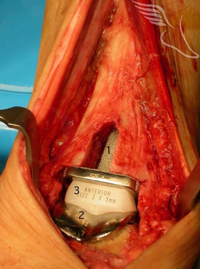
The “eureka” moment in ankle replacement came with the introduction of 3 component replacements in which the Tibial and Talar components were linked by a UHMW polyethylene meniscus which allowed an element of rotation to occur within the joint itself as well as providing adequate component stability.
These initial replacements whose results still define what longevity an ankle replacement should attain are the STAR , Beuchal-Pappas and Salto implants. In general their 10 year survivorships are lower than reported for hip and knee replacements but are still in the mid to high 80 percents.
In general terms an ankle replacement will not be recommended for younger and higher demand patients due to concerns of accelerated wear of the implant .

INDICATIONS
–Isolated Ankle arthritis in a well aligned ankle: Generally a replacement is operation for the lower demand and fifty plus age group with limited angular deformity and good soft tissue cover around the ankle and with no history of deep infection or neuropathy. With an ankle replacement the failure rate of most implants (which have been in use for long enough) is 2%/annum which equates to a 10 year survivorship of 80%.
A fusion in general is for higher demand/ younger patients or those wishing a greater degree of predictability than afforded by Ankle replacement. With a fusion the “risk” in the majority of patients can be regarded as “front-loaded”. As long as a non-union does not occur (5-10% chance, technique dependent) then in the majority no subsequent / later intervention is likely though the subtalar and midfoot joints are highly likely to become degenerate. Function will reduce with this if this occurs but the lead time is likely to be 10-20 years. .
-Ankle arthritis with deformity : This will self evidently be a more challenging primary operation. In cases of deformity with bone loss effecting the weight bearing surfaces then cutting the deformity out of the bone may lead to large bony resections .This can produce issues with adequate bony support for the prosthesis and potentially relative laxity of the soft tissues stabilisers . The context most often is a varus arthritic ankle where a robust lateral ligament reconstruction may be required at the time of primary operation (such as an Evans peroneal re-routing stabilisation).
Relative indications for ankle replacement (when compared to Ankle Fusion) are also for cases of inflammatory arthropathies involving the ankle, where subtalar and mid foot articulations have (or are more likely to develop) inter-current disease and on one side for a patient with bilateral ankle arthritis.
SYMPTOMS & EXAMINATION:
Most patients with severe ankle arthritis localise the pain well to the level of the joint. Very much as with arthritis elsewhere symptoms tend to progress from early activity /start up pain which eases off through to progressively more disabling and continual weight bearing pain and on occasion as far as pain at night or at rest. A much less common symptom which can co-exist with pain is that of ankle instability. If gait is becoming altered due to the arthritis pain proximal to the ankle may occur secondary to alteration of the weight-bearing axis of the limb.
The vast majority of patients will either have a history of a significant injury (such as an ankle fracture), chronic deformity (for example Cavo-varus) or a past history of chronic lateral ligament instability. More rarely the cause is a more generalised tendency to osteoarthritis or an inflammatory arthropathy.
On examination swelling and tenderness well localised to the ankle is common. Range of movement is often reduced and may be uncomfortable. More important than ankle movement is what the subtalar and midfoot mobility is like. If both are very mobile then it is likely that post-fusion good compensatory movement in these joints will allow normal gait and in fitter ,younger patients even the ability to return to running. Conversely if movement here is restricted these joints should be carefully inspected with CT to confirm or refute additional arthritic change. If still equivocal then an injection into the ankle joint with inta-articular contrast (see below) is indicated.
Any deformity should be noted. Varus is most common and valgus and equinus less common. The key issues with any deformity are A:Whether it is passively correctable (or not) and B.:Being sure of its anatomical location(s). The former is easily clinically determined .The latter can be more difficult to be sure on , in particular in the presence of severe deformity and CT is indicated for this.
Another feature to examine carefully in any varus ankle is the position of the 1st Ray , in particular whether it is plantar flexed and fixed. If this becomes apparent with the ankle corrected to neutral then the first ray should be inter-currently corrected .
In assessing equinus it should be appreciated at what level(s) the deformity rests. Beware of associated fixed midfoot equinus which will leave the mid/forefoot in a plantar flexed position once the ankle is fused in neutral if it is ignored. A midpoint plantar fascia release may be all that is required to place the foot in a functional position post-operatively. If dealing with isolated ankle equinus be prepared to add a triple cut (or open )Achilles release dependent on the severity of the deformity.
The rest of the lower limbs alignment should not be forgotten. In general correction of deformity should start proximally and proceed distally.
A vascular examination must be made and if abnormal dealt with appropriately.
INVESTIGATION:
Plain X-Ray: This is the initial imaging for most patients with ankle arthritis of any degree. Though the ankle is relatively well visualised (and the films should be taken weight-bearing) the subtalar and midfoot joints aren’t so well shown , in particular in the presence of associated deformity through the area.
CT scan. This is better in defining how much relevant arthritic change exists and where it is than MRI. It is also easier to differentiate the level of deformity from CT than MRI. There are cases where significant cystic change exists and will require bone grafting.Its location and extent is again best defined with CT.
MRI scan: An MRI is more sensitive for early degenerative change but will be degraded by any internal fixation and is not 100% sensitive for early arthritis. It can be more difficult to be objective about the severity of more advanced arthritic change as bone oedema ( a reversible phenomenum) complicates the MRI images. A CT lacks this sensitivity which is a positive and not a negative. Some surgeons prefer to use MRI rather than CT pre fusion as imaging.
X-Ray guided injection: This should be into whichever joint (ankle or subtalar ) appears more likely the location of symptoms. Contrast is needed as in a proportion of patients the two joints will inter-connect and improvement of symptoms after injection into one cannot under these circumstances be regarded as discriminatory.
ALTERNATE OPERATIVE MANAGEMENT:
Open Ankle fusion:The union rates are continually demonstrated to be the highest with the arthroscopic technique , pain levels are lower than for open surgery and hospital stay in half of patients is just one night. Ultimately no longer term difference with a successful Arthroscopic versus Open ankle fusion , just more patients get there arthroscopically and the journey is easier. In the presence of severe and fixed deformity however it should be the procedure of choice.
Arthroscopic Ankle debridement: This has a role for the treatment of those with lesser degrees of arthritic change & “intermediate” symptoms. There are no clear criteria for this but patients with severe levels of pain on minor activity and possibly at rest or night are unlikely to be appropriate candidates for joint sparing surgery.
Arthroscopic Ankle Fusion:
Distal Tibial osteotomy:
NON-OPERATIVE MANAGEMENT:
Activity modification and analgesia.
Local anaesthetic & steroid injection.
Orthotics & Shoewear modifications.
CONTRAINDICATIONS:
Active infection , active smoking ,poor vascular inflow: require correction before replacement is considered.

GA or regional anaesthesia
Femoral & sciatic blocks for post-operative pain relief
Laminar flow , peri-operative antibiotics , 2-4 weeks of post operative LMW Heparin
Thigh tourniquet and Flowtron on contra-lateral calf
Supine position
Large , rolled up sterile towels behind the ankle to ease positioning & movement of the joint .


How much weight bearing allowed early depends upon the quality of the press fit of components and a Surgeons preference. My own is to limit al weight-bearing for at least 4 weeks with a light weigh cast.This does not have the disastrous effect upon range of movement ( or indeed I think any effect)that one would expect if treating a knee replacement in a similar fashion.The incidence of early post-operative peri-articular fracture is significantly reduced.
Once out of cast follow with a post-operative boot boot , week 7-12 .
Range of movement can be worked upon once anterior wound has healed.
Post operative follow up xrays weeks 6,12 and annually.

Total Ankle Replacement.The results in 200 Ankles.
P.L.R.Wood , S.Deakin .
J Bone Joint Surg(Br).2003;85-B:334-41.
200 uncemented replacements. Mean follow up 46 months. 5 year survival 92.7%
5 major wound problems, 7 ongoing pain & stiffness ,19 fractured malleoli .
A randomised , controlled trial of two mobile bearing total ankle replacements.
P.L.R.Wood, C.Sutton , V. Mishra, R.Suneja
J Bone Joint Surg(Br).2009;91-B:69-74.
RCT of STAR versus Beuchel-Pappas implants. 6 year survivorship 79% BP and STAR 95%.
Not however a statistically significant difference.
Kinematics of a Total Arthroplasty of the ankle:Comparison to Normal ankle motion.
J.D.Michelson, G.R.Schmidt,M.S.Mizel
Foot & Ankle International.Vol.21 ,No 4:
278-284
The management of failed ankle replacement.
J Bone Joint Surg.2006. 88-B:1039-1047.
K.Kotnis , C.Pasapula , F.Anwar , P.H.Cooke ,R.J.Sharpe.
Reference
- orthoracle.com


