
Learn the Closed reduction and K wiring of 5th carpometacarpal fracture-dislocation surgical technique with step by step instructions on OrthOracle. Our e-learning platform contains high resolution images and a certified CME of the Closed reduction and K wiring of 5th carpometacarpal fracture-dislocation surgical procedure.
A fracture dislocation of the 5th carpo-metacarpal joint is the second most common injury in the hand, after the thumb carpo-meatcarpal joint. The injury is caused by an axial loading force, most commonly seen in punching injuries. Given this they are common in adult males of a younger age group, may present with associated dirty puncture wounds to the metacarpo-phalangeal joints and may present with or without associated fracture. Most often they are closed injuries and require reduction and stabilisation.
An axial loading force to a clenched fist causes these injuries. The loading vector often results in a metacarpal neck fracture. The force can alternatively be transmitted proximally at the CMC joint resulting in its fracture-dislocation. The bases of the 4th and 5th metacarpals articulate with the hamate and are stabilized by dorsal and palmar intermetacarpal ligaments along with the interosseous ligament. The deforming force causes the metacarpal base to subluxate or dislocate with or without an avulsion fragment from the hamate. Occasionally, there may be an associated fracture involving the base of the metacarpus. The radial 25% of the metacarpal base may remain intact in its normal anatomical location. In this respect, this injury is not dissimilar to a Bennet’s fracture at the base of the thumb. Once displaced, the deformity is accentuated by the pull of the extensor and flexor carpi ulnaris as well as the abductor digiti minimi.

INDICATIONS:
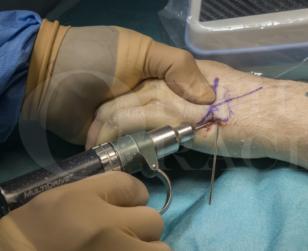
Subluxation or dislocation of a joint should always be reduced and stabilized. Manipulation and percutaneous K wiring is my method of choice in juries with pure dislocation or those with minor associated fractures of the Hamate (Type 1 injuries and some Type 2)
SYMPTOMS & EXAMINATION
Patients present with pain and swelling over the dorso-ulnar border of the hand. There is tenderness over the base of the metacarpus and movements of the finger may be restricted with pain. A common finding is the presence of a bony lump over the base of the metacarpus. There may be an apparent extensor lag of the small finger at the metacarpophalangeal joint. Presence of any rotational abnormality should be evaluated and documented. Comparison can be made with the opposite uninjured hand.
IMAGING
Plain radiographs will confirm the diagnosis and aid in planning the management. Always request three radiographic views of the hand– Anteroposterior, lateral and oblique. The lateral radiographic view is essential to identify the dorsal subluxation/dislocation of the base of the metacarpus. It is not uncommon to miss this in the routine AP and oblique radiographs. The metacarpal height is reduced and the metacarpal head will appear to be flexed forward. Any concomitant fractures of the base are identified and their intra-articular extensions documented. An associated fracture of the dorsal lip of hamate should be looked for.
Cain, Shepler & Wilson graded these injuries into 3 types depending upon the fracture pattern of the hamate in relation to the CMCJ subluxation.
Type IA lesions are characterized by subluxation or dislocation of the fifth carpometacarpal joint without hamate avulsion fracture.
Type IB lesions are identical to type IA lesions except for the appearance of a small dorsal rim hamate avulsion fracture.
Type II lesions are distinguished by subluxation or dislocation of the fifth carpometacarpal joint and comminution of the dorsal hamate rim.
Type III lesions exhibit coronal splitting of the hamate.
A CT scan is useful in identifying the various fracture fragments and their alignment. This is especially useful in delineating coronal splits in the hamate to guide internal fixation.
ALTERNATIVE OPERATIVE TREATMENT:
Masterful inactivity – Immediate mobilization of the hand and fingers with pain relief has been tried by Petrie & Lamb. Despite obvious radiographic abnormalities at 4-year follow-up, there was no significant functional disability identified in their cohort. These results have not been reproducible elsewhere.
Conservative with closed reduction followed by plaster immobilisation – A plaster cast or splint is often inadequate to maintain reduction as the injury is fairly unstable due to the forces of the Extensor and Flexor carpi ulnaris and Abductor Digiti Minimi. If treated conservatively, regular radiographs at weekly intervals are necessary to identify any redisplacement.
Closed reduction and external fixator – This device will span the CMC joint and allows reduction with ligamentotaxis. Placement of proximal pins in the small carpal bones can be technically challenging. In addition, the device requires meticulous pin site care in the postoperative period to prevent infections and complications.
Open reduction and internal fixation with plates and screws – This can provide rigid stabilization and early mobilization of the hand. However, there is a clear risk of injury to the dorsal branch of ulnar nerve during the procedure. Tendon adhesions remain a significant concern. Moreover, the comminution of fracture fragments may preclude satisfactory anatomical extra-articular stabilization necessitating a bridge plate across the CMC joint and a secondary procedure for removal of metalwork. In summary, this remains the treatment modality of choice in injuries that are more than 3 weeks old where closed reduction with manipulation is likely to fail as well as those with associated comminuted fractures.
Salvage – Untreated or redisplaced injuries often present with pain and reduced grip strength. This is related to secondary osteoarthritis at the CMC joint with or without persistent subluxation. Such cases will require salvage procedures such as resection arthroplasty, interposition arthroplasty or an arthrodesis.
CONTRAINDICATIONS:
There are no specific contraindications to this procedure. A note of caution needs to be exercised in injuries with associated comminuted fractures of the base of the metacarpus or of the hamate – as these may not be adequately stabilised with K-wires.
Cain JE, Shepler TR, Wilson MR. Hamatometacarpal fracture-dislocation: classification and treatment. Journal of Hand Surgery. 1987 Sep 1;12(5):762-7.

Informed consent is an important part of the procedure and the risks and benefits should be clearly explained to the patient. The dorsal branch of ulnar nerve lies in close proximity and can be injured either by the fracture fragments or the percutaneous (and therefore blind) pin insertion. The intra-articular fragmentation at the base of the metacarpal can be a precursor for future secondary osteoarthritis of the CMC joint.
I prefer regional anaesthesia with axillary block for this procedure. The patient is placed supine with the limb extended on an arm table. Upper arm tourniquet is applied and inflated after exsanguination. A prescrub is performed followed by a sterile prep with Chlorhexidine. A lead hand is often not required for this procedure. I routinely administer a single dose of antibiotics for this procedure.


Finger mobilization exercises are commenced immediately. The wound is redressed and the plaster cast changed at 2 weeks. There are no sutures to remove.
The plaster is discarded and radiographs repeated at 4-6 weeks. K wires are removed in the clinic at this stage. Full mobilisation exercises of the hand and wrist can now be instituted and progressively increased. Activities of daily living can be commenced at this stage. I advise patients against heavy activities and manual labour for atleast 3 months – until the fracture is consolidated. I have rarely needed to repeat radiographs after 6 weeks.
Pintrack infections should be watched for and treated accordingly. Comorbidities that compromise the immune status of the patient are more likely to present with superficial or deep infections following the procedure. Superficial cellulitis can be managed with wound care and oral antibiotics. I have rarely needed to remove the K wires early due to purulent discharge at the pinsite. These patients have their hands protected in a plaster cast following the early removal of metalwork for a total of 6 weeks following the initial surgical intervention. I have not encountered recurrent subluxation of the CMC joint in any of these cases, although they have been reported in literature and may be related to inadequate stabilisation.
Some reduction in grip strength is commonly noted and reported in literature. Incidence of secondary osteoarthritis is related to the severity of the fracture comminution. However, as the CMC joint has very limited mobility, these rarely require further intervention.

Cobb WA, Dingle L, Zarb Adami R, Rodrigues J. Management of fracture-dislocations of the little finger carpometacarpal joint: a systematic review. Journal of Hand Surgery (European Volume). 2018 Jun;43(5):530-8.
This is a recent systematic review evaluating the effectiveness of all the various modalities of treatment for these injuries. Unfortunately, due to the heterogenicity of the data, the authors were unable to identify a single best modality. They suggest to plan treatment on an individual case basis. The article is a rich source of references for the management of this condition.
Cain JE, Shepler TR, Wilson MR. Hamatometacarpal fracture-dislocation: classification and treatment. Journal of Hand Surgery. 1987 Sep 1;12(5):762-7.
Although this is a series of just 17 patients, the authors have recognised different injury patterns and have proposed a simple but effective classification of these injuries. The classification is very useful in planning treatment and I feel that Percutaneous K-wiring can be used in type1 and type 2 injuries. Type 3 injuries will require internal fixation with plates/screws
Frick L, Mezzadri G, Yzem I, Plotard F, Herzberg G. Acute carpometacarpal joint dislocation of the long fingers: study of 100 cases. Chirurgie de la main. 2011 Oct;30(5):333-9.
This is one of the largest case series in literature looking at 100 patients with this injury. The authors report an increased incidence of loss of reduction with closed reduction and percutaneous wiring as compared to open reduction. However, this has not been my experience using 2 K-wires in crossed configuration.
Reference
- orthoracle.com


